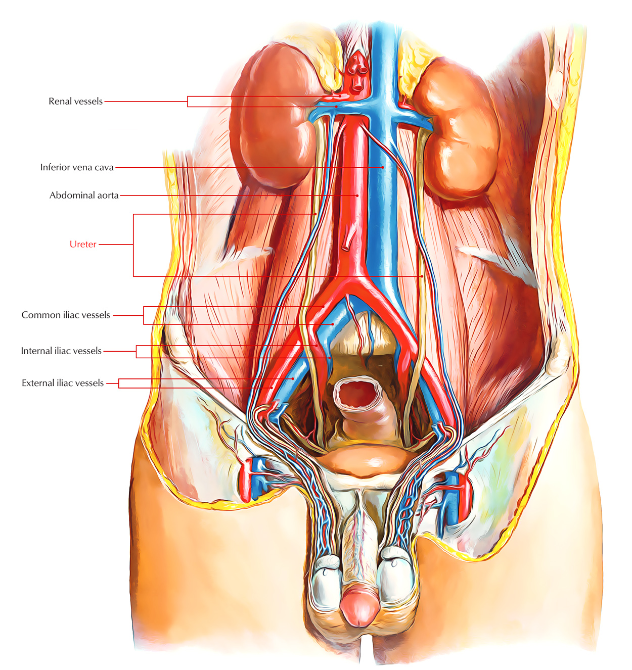
The Ureter is a narrow, thick-walled, expansile muscular tube that carries the urine from the kidney to the urinary bladder. By the peristaltic contractions of the smooth muscle of the wall of the ureter, the urine is propelled from the kidney to the urinary bladder.
Ureter
Measurements
- Length: 25 cm (10 inches).
- Diameter: 3 mm.
Course
- The ureter is connected as a downward continuance to the renal pelvis, which is a funnel-shaped organ, to the medial margin of the lower end of the kidney.
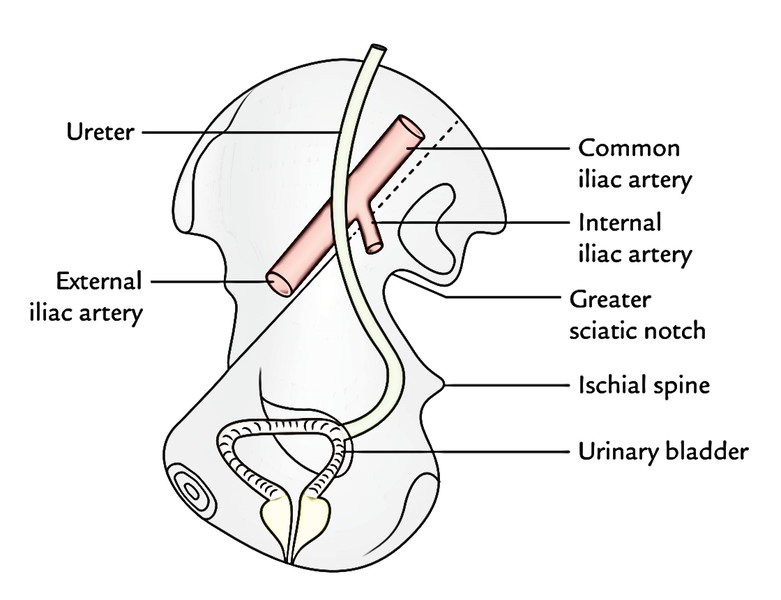
- The end of the ureter is a little medially on the psoas major and then it enters downwards. This process divides it from the traverse of the lumbar vertebrae and enters the pelvic cavity by crossing in front of the bifurcation of the common iliac artery in the pelvic brim in front of the sacroiliac joint.
- In the pelvis, the ureter first runs downward, backward, and laterally along the anterior margin of the greater sciatic notch. Opposite to the ischial spine, it turns forwards and medially to get to the base of the urinary bladder, where it enters the bladder wall obliquely. Inside the bladder wall, it narrows down, takes a sinuous course, and opens into the cavity of the bladder in the lateral angle of its trigone as ureteric orifice.
Ureter: Course
- During its course from the kidney to the urinary bladder, it runs behind the parietal peritoneum to which it’s closely implemented.
- The renal pelvis is an upward funnel-shaped continuance of the ureter and thus also referred to as pelvis of the ureter. It is located partially outside and partly inside the kidney.
Parts and Relations
- The ureter is normally split into 2 parts: abdominal and pelvic. Every part is all about precisely the same length, i.e., 12.5 cm (5 inches).
- The abdominal part of ureter goes from the renal pelvis to the bifurcation of the common iliac artery.
- The pelvic part of the ureter goes from the pelvic brim (at the level of bifurcation of the common iliac artery) to the base of the urinary bladder.
Abdominal Part
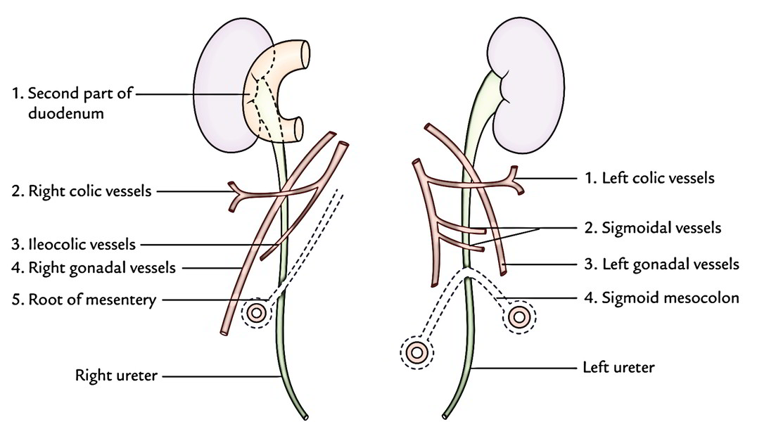
The anterior and posterior relationships of the abdominal part of the ureter are provided in Table below.
Ureter: Abdominal Part
Medially the right ureter is related to inferior vena cava and left ureter is related to left gonadal vein and inferior mesenteric vein.
Pelvic Part
The pelvic part of the ureter crosses in front of all the nerves and vessels on the lateral pelvic wall with the exception of vas deferens, which crosses in front of it. Near the uterine cervix, the uterine artery is located above and in front of it, an extremely essential surgical association.
| Anterior relations | Posterior relations | ||
| Right ureter |
|
| |
| Left ureter |
|
| |
Sites of Anatomical Narrowings/ Constrictions
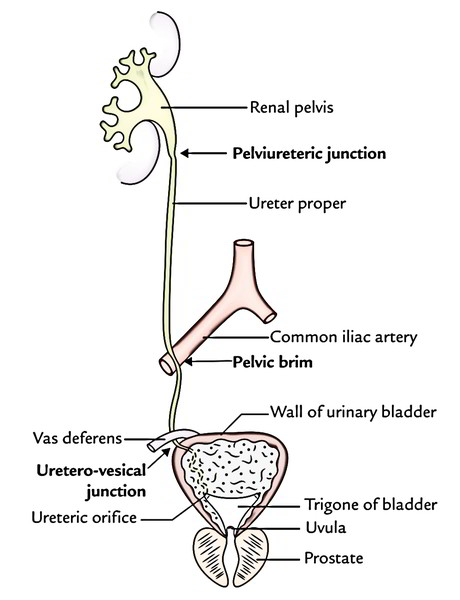
The lumen of the ureter isn’t consistent throughout and presents 3 constrictions at these sites:
- At the pelviureteric junction where the renal pelvis joins the upper end of ureter.
It’s the upper most constriction, seen roughly 5 cm far from the hilum of kidney.
- At the pelvic brim where it crosses the common iliac artery.
- At the uretero-vesical junction (i.e., where ureter enters into the bladder).
Ureter: Site of Anatomical Narrowings
Portions of the ureter between these constrictions reveal spindle shaped dilatations. These constricted parts of the ureter are the sites of arrest of ureteric calculi.
In addition to above 3 sites of constrictions, 2 more sites of constrictions are described by the surgeons, 1 at juxtaposition of the vas deferens/broad ligament and other at the ureteric orifice.
Arterial Supply
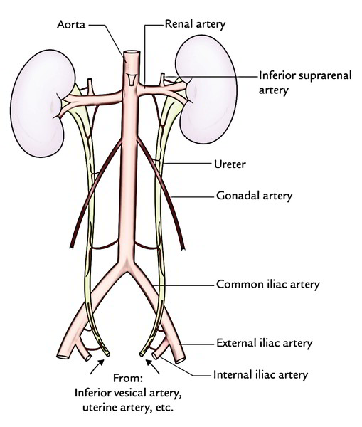
The ureter derives its arterial supply from the branches of all the arteries related to it. The significant arteries providing ureter from above downwards are:
- Renal.
- Testicular or ovarian.
- Direct branches from aorta.
- Internal iliac.
- Vesical (superior and inferior).
- Middle rectal.
- Uterine.
Ureter: Arterial Supply
Arteries supplying the ureter split into ascending and descending branches to first create a plexus in the connective tissue sheath on the top layer of the ureter and after that supply it.
Venous Drainage
The venous blood from the ureter is drained into the veins corresponding to the arteries.
Lymphatic Drainage
The lymph from the ureter is drained into lateral aortic and iliac nodes.
Nerve Supply
- The sympathetic supply of the ureter is originated from T12 L1 spinal sections via renal, aortic, and hypogastric plexuses.
- The parasympathetic supply of ureter is originated from S2 S4 spinal sections via pelvic splanchnic nerves.
The afferent fibres go with both sympathetic and parasympathetic nerves.
Clinical Significance
Mobilization of Ureter
Branches of the arteries supplying the ureter create an anastomosis in the fat and fascia around the ureter. Consequently, surgeons should endure in their own head that stripping off this fascia, while marshalling the ureter for transplantation, will hamper the blood supply of the ureter and could cause its necrosis.
Identification of Ureter
Ureter is a muscular structure, and in life waves of muscular contractions generate a worm like rhythmic movement (peristalsis) consequently milking pee toward the bladder. The ureter is easily identified in life by its thick muscular wall that is observed to go through worm like writhing movements, notably when it’s lightly stroked or squeezed. Violent muscular contractions precipitated by the presence of stone in the lumen of the ureter (ureteric calculus) create this kind of acute spasmodic pain referred to as renal colic that immediate treatment is needed.
Ureteric Colic
It takes place because of blockage of ureteric lumen by a stone. The referred pain of ureteric colic is associated with the cutaneous regions innervated by exactly the same spinal segments as that of the ureter, i.e., T12 L2. Pain of ureteric colic commences in the loin, shoots downward and forward to the groin and after that into the scrotum or labium majus.
- Pain from upper ureteral obstruction is referred to the lumbar region (T12 and L1).
- Pain from middle ureteral obstruction is referred to the inguinal, scrotal or mons pubis, and upper medial aspect of the thigh (L1, L2).
- Pain from lower ureteral obstruction is referred to the perineum (S2 S4).
Localization of a Ureteric Stone on the Plain Radiograph of the Abdomen
To localize the stone in the ureter in plane X-ray abdomen, one must understand the course of ureter in connection to the bony skeleton. Ureter is located in front of the tips of the transverse processes of the lower 4 lumbar vertebrae, crosses in front of the sacroiliac joint, swings out to the spine of the ischium, and after that runs medially to the urinary bladder. In plane X-ray of abdomen, for that reason the radiopaque shadow of ureteric calculus is generally viewed in these sites:
- Near the tips of the transverse processes of lumbar vertebrae.
- Overlying the sacroiliac joint.
- Overlying or somewhat medial to the ischial spine.
Injury to Ureter
Based on Kenson and Hinman, the ureter could possibly be injured at one of many subsequent 4 dangerous sites:
- Point where the ureter crosses the iliac vessels.
- In the ovarian fossa.
- Where the ureter is crossed by the uterine artery (most dangerous site) as damage is likely at this site during hysterectomy.
- At the base of the bladder.
Ureteric Calculus
Ureteric calculus probably will stay at 1 of the sites of anatomical narrowings of the ureter especially:
- At the pelvic ureteric junction.
- Where it crosses the pelvic brim.
- In the intramural part- the narrowest part.
Approach to Ureter
Throughout its abdominal and upper parts of the pelvic course, the ureter runs deep to the peritoneum and sticks with it closely. During surgery when the ureter is mobilized, the ureter is in danger of injury for it moves with the peritoneum. Deep to the peritoneum in the abdominal part the ureter is crossed by different blood vessels. Because of these vascular Relations an extraperitoneal method of the ureter is favored to that of a transperitoneal method.
Development
The ureter grows from the ureteric bud appearing as an outgrowth from the mesonephric duct.
Clinical Significance
Congenital anomalies:
- The common congenital anomalies of ureter are: (a) double pelvis, (b) bifid ureter, and (c) double ureters (upper ureter enters below the lower ureter). Seldom the additional ureter may open ectopically into the urethra or vagina and cause urinary incontinence.
- The cause of double pelvis is early division of the ureteric bud near its conclusion, on the other hand the cause of bifid or double ureter is the overly early section of the ureteric bud.
- Occasionally during the rise of kidney, the ureter may ascend posterior to the inferior vena cava resulting in postcaval ureter.

 (60 votes, average: 4.75 out of 5)