Urethra, a tabular passage, is the organ of the body which carries urine and seminal fluid in men and only urine in females. The essential medical processes such as catheterization and cystoscopy can only be performed after the study of urethra. One of the common phenomenon is urethral rupture.
Male Urethra

Urethra: Male Urethra
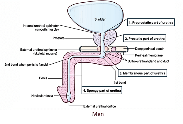
The length of a male urethra is about 18-20 cm.
It stretches from the internal urethral orifice in the neck of the urinary bladder to the external urethral orifice (EUO) at the tip of the glans penis.
In flaccid state of the penis, the long axis of the urethra presents 2 curvatures and is consequently S-shaped. In erect state of the penis, the distal curvature goes away and consequently, it becomes ‘J-shaped’.
Parts
According to its location, the urethra is split into the following 3 parts:
- Prostatic part (goes through the prostate).
- Membranous part (goes through the urogenital diaphragm).
- Spongy or penile part (goes through the corpus spongiosum of penis).
Prostatic Part of The Urethra (3 Cm Long)

Urethra: Parts of Urethra
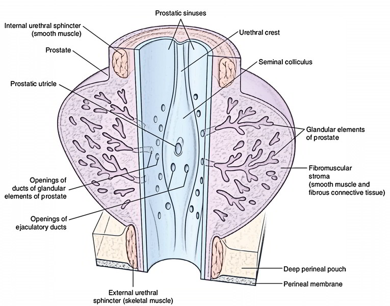
As its name suggests it traverses via the anterior part of the prostate. It’s the broadest and most dilatable part of the male urethra. It’s fusiform in the coronal section. The inner aspect of its posterior wall presents these features:
- Urethral crest, a median longitudinal ridge of the mucous membrane.
- Colliculus seminalis (verumontanum), an elevation on the middle of the urethral crest. The prostatic utricle opens on its peak by a slit-like orifice. On each side of the orifice of the prostatic utricle opens the ejaculatory ducts.
- Prostatic sinuses, vertical grooves 1 on every side of the urethral crest. Every sinus presents 15-20 openings of the prostatic glands.
Prostatic utricle: It’s a mucous culdesac (5.5 cm long) in the substance of the median lobe of the prostate. It develops from united caudal ends of the 2 Mullerian ducts; thus it corresponds to (i.e., homologous to) the uterus and vagina of the female. It’s also called vaginus masculinus.
Membranous Part of The Urethra (2 Cm Long)
It traverses via the urogenital diaphragm and pierces the perineal membrane about 2.5 cm below and behind the pubic symphysis. It’s encompassed by the sphincter urethrae muscle, which acts as the voluntary external sphincter of the bladder.
With the exclusion of external urethral orifice, it’s the narrowest and least dilatable part of the urethra. Numerous mucous glands are frequently seen in it. In cross section, its lumen is star shaped.
Spongy Part of the Urethra (15 Cm Long)
It traverses via the corpus spongiosum of the penis. It first enters upward and forwards in the bulb of penis to be located below the pubic symphysis. Subsequently it bends downward and forward, and traverses the corpus spongiosum in the free part of the penis and ends as the external urethral orifice just below the tip of glans penis.
It presents 2 dilatations: (a) in the bulb of penis to create intrabulbar fossa (3 cm long) and (b) in the glans penis to create navicular fossa/terminal fossa (1.25 cm long).
In cross section, the shape of the spongy urethra differs in various parts, viz., trapezoid-shaped in the bulb, like a transverse slit in the body and like a vertical slit at the external urethral orifice.
- The small simple tubular mucous glands named urethral glands (Littre’s glands) open in the whole spongy part of the urethra with the exception of in the terminal fossa.
- The pit-like small mucous recesses, the urethral lacunae (of Morgagni) project from the whole spongy part of the urethra with the exception of in the terminal fossa. The lacunae get the openings of urethral glands. 1 lacuna within the roof of terminal fossa is referred to as lacuna magna or sinus of Guerin.
The external urethral orifice is the narrowest part of the male urethra. It’s in the form of a sagittal slit about 6 millimeters long.
Distinct Parts of the Male Urethra
| Features | Prostatic part | Membranous part | Spongy part |
|---|---|---|---|
| Location | Within prostate | Within urogenital diaphragm | Within penis (corpus spongiosum) |
| Length | 3 cm | 1.5-2 cm | 15 cm |
| Shape in cross section | Horseshoe shaped | Star-shaped | In the bulb—trapezoid |
| In the body—transverse slit | In the body—transverse slit | In the body—transverse slit | In the body—transverse slit |
| At external urethral orifice—vertical slit | At external urethral orifice—vertical slit | At external urethral orifice—vertical slit | At external urethral orifice—vertical slit |
| Diameter and dilatability | Widest and most dilatable | Narrowest and least dilatable | Mostly uniform diameter with medium dilatation |
| Openings | 1. Prostatic utricle 2. Ejaculatory ducts 3. Prostatic glands | Minute mucous glands | Urethral glands (Littre’s glands) |
The lumen of the male urethra is unusual, i.e., it’s different shapes in different parts. This makes the urine projectile in nature and gives a coil wind to the urinary flow. Consequently, the early separation of droplets of urine doesn’t happen which prevents the wetting of the garments.
Urethral Mucosa
The urethral mucosa presents regional variations as follows:
- Prostatic urethra above the seminal colliculus is lined by transitional epithelium and below it by stratified columnar epithelium.
- Membranous urethra is lined by stratified columnar epithelium.
- Spongy urethra up to navicular fossa is lined by stratified columnar epithelium. The navicular fossa and external urethral orifice are lined by stratified squamous epithelium.
Clinical Significance
Rupture of the Urethra
The rupture of the urethra contributes to extravasation of urine. The commonest site of rupture is bulb of the penis, just below the urogenital diaphragm following a fall astride a sharp object. The urethra is squashed against the border of the pubic bones. The urine extravasates into the superficial perineal pouch and enters forward over the scrotum, penis, and anterior part of the anterior abdominal wall deep to membranous layer of the superficial fascia (superficial extravasation). If the urethra ruptures above urogenital diaphragm urine escapes above the deep perineal pouch and can pass upward around the prostate and bladder in the extraperitoneal space (deep extravasation).
Catheterization of the Male Urethra
It’s done in the patients that can not pass urine and their bladders are distended because of retention of urine resulting in serious distress and pain in the hypogastric region. While passing the catheter one should remember the normal curvatures of the urethra. Additional one should understand that immediately above the external urethral meatus the urethra presents a large mucosal recess safeguarded by a mucosal fold, which might catch the tip of catheter. The catheter/iron bougie should thus constantly be added into the urethra with its beak turned downward. Otherwise, the strong insertion of catheter may produce a fake passage in urethra or rupture it.
Hypospadias
It’s a congenital anomaly where external urethral orifice lies on the inferior/ventral aspect of penis instead at the tip of the glans penis. It happens because of failure of the fusion of urethral folds. Contingent on the location of external urethral orifice (EUO), the hypospadias are classified into 5 types:
- Glandular: If external urethral orifice opens on the under surface of the glans.
- Balanic: If urethral orifice opens at the base of glans penis.
- Penile: If urethral orifice opens on ventral aspect of the body of penis.
- Penioscrotal: If urethral orifice opens at the junction of penis and scrotum.
- Perineal: If urethral orifice opens as sagittal slit on ventral aspect of scrotum.
Sphincters of the Urethra
- The urethra has 2 sphincters- internal and external.
Internal Sphincter
The internal sphincter encircles the internal urethral orifice and is likely created from the muscle of the bladder wall. It’s automatic in nature and frequently named sphincter vesicae. It’s supplied by the sympathetic fibres from lower thoracic and upper lumbar segments of the spinal cord (T11 to L2). It relaxes during urination but shuts (i.e., contracts) during ejaculation (to prevent the retrograde entrance of semen into the bladder).
External Sphincter (or Sphincter Urethrae)
The external sphincter encircles the membranous part of the urethra and is originated from the sphincter urethrae muscle. It’s voluntary in nature and is furnished by the pudendal nerve (S2, S3, S4).
Differences Between the Internal and External Sphincters of the Urethra
| Internal urethral sphincter | External urethral sphincter |
|---|---|
| Surrounds the internal | Surrounds the membranous |
| urethral orifice | part of urethra |
| Derived from the bladder | Derived from the sphincter |
| musculature of trigonal region | urethrae muscle |
| Innervated by the | Innervated by the somatic |
| sympathetic fibres (T11-L2 segments) | fibres (S2, S3, S4 segments) |
| Involuntary | Voluntary |
Female Urethra

Urethra: Female Urethra
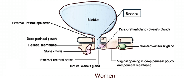
The female urethra is all about 4 cm long. It starts at the internal urethral orifice in the neck of bladder and enters downward and forwards embedded in the anterior wall of the vagina via urogenital diaphragm. Urethra pierces the perineal membrane, and opens in the vestibule of vagina in front of the vaginal orifice. In the vestibule of vagina, the urethral orifice is situated in front of the vaginal orifice and about 2.5 cm behind the glans of clitoris.
Shape of the Female Urethra
In cross section, the shape of female urethra differs in various parts:
- In the upper part, it’s crescentic with convexity directed forwards.
- In the middle part, it’s star-shaped (stellate-shaped).
- In the lower part, it’s a transverse slit.
- At the external urethral orifice, it’s a sagittal slit.
Glands and Lacunae Around the Female Urethra
- Urethral glands: All these are small tubular glands and surround the complete urethra.
- Paraurethral glands (of Skene): All these are comparatively large mucous glands and aggregated on every side of the upper part of the urethra. These glands are homologous to the male prostate.
- Urethral lacunae: All these are pit-like mucous recesses along the whole urethra.
Clinical Significance
- The female urethra is easily dilatable, thus catheter/ cystoscope can be easily managed.
- Urinary tract infections are far more common in females because of shortness of the urethra and presence of its orifice on the surface near the vaginal and anal orifices. So, females must take bath at least once per day.
- Urinary incontinence is common in females because in them external urethral sphincter is a tenuous structure and is further weakened during childbirth.

 (48 votes, average: 4.56 out of 5)