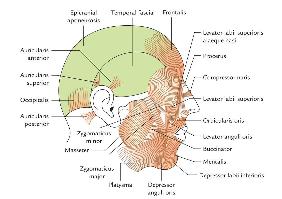
The zygomaticus is a set of small muscles which travel via eye socket till the lips, beside the cheekbone. They attach with the zygomatic bone a.k.a. the cheekbone.
The zygomaticus major directs the motion of upper lip outward and superiorly, and it especially controls smiling. It is activated when one frowns or shows sadness. The zygomaticus minor is located anterior towards the zygomaticus major as well as supports the larger muscle in order to shifts the upper lip upwards and outwards and backward as well.

Zygomaticus Major and Minor
Zygomaticus Major
Origin
The zygomaticus major originates deep towards the orbicularis oculi together with the posterior portion of the lateral side of the zygomatic bone, and afterwards it passes inferiorly and anteriorly, merging together with the orbicularis oris and attaching at the angle of the mouth within the skin.
Insertion
It attaches on the angle of the mouth in the muscular node. Nearby the node, a number of inserting fibers may mix in the risorius or the depressor anguli oris. There are rare attachments within the skin of the lower end of the nasolabial furrow.
Structure
The zygomaticus major is a long, slim muscular strap, due to its length; widespread shifting of the node as well as angle of the mouth can be done. The zygomaticus major divides within superficial as well as deep portions while it extends to its insertion. Frequently, the superficial part divides within two narrow bellies.
Innervation
The buccal and zygomatic branches of the facial nerve (7th cranial nerve) innervate the zygomatic major.
Artery Supply
Facial artery supplies both zygomaticus major and minor muscles.
Action
The zygomaticus major draws the complete lower face outward as well as upward. The entire cheek rises and protrudes. The lower eyelid shifts slightly over the iris. The wrinkles inferior to the eye develop, and at the outer angle of the eye crow’s feet develop. The orbicularis oculi helps these effects because it frequently shrinks at the time of strong narrowing of the zygomaticus major.
Zygomaticus Minor
The zygomaticus minor originates via the zygomatic bone anterior towards the starting point of the zygomaticus major, matches the path of the zygomaticus major, and attaches medial towards the corner of the mouth within the upper lip. Both zygomaticus muscles elevate the corner of the mouth as well as move it lateralward.
Origin
Zygomaticus minor originates via the anterior side of the zygomatic bone, inferior towards the lateral margin of the orbit.
Insertion
It attaches to the skin of the middle section of the nasolabial furrow as well as into the cheek fat. Other fibers extend inferiorly further towards the red lip, traveling over and across the mass of the orbicularis oris.
Structure
From its origin, it travels medially and then arcs inferiorly. At its origin, it is located deep towards the orbicularis oculi.
Action
It draws the middle portion of the nasolabial furrow along with the middle portion of one side of the upper lip outward as well as slightly upward. The angle of the mouth is not drawn by the zygomaticus minor.
Innervation
Zygomaticus minor is innervated by the facial nerve (CN VII).
Artery Supply
Facial artery supplies both zygomaticus major and minor muscles.
Clinical Significance
Temporal Tendinitis
Temporal tendinitis is an inflammation of the tendon which goes via the temple till the jaw. This inflammation creates:
- Pain in the zygomaticus zone
- Headache
- Ear pain
- Soreness in the Jaw
- Eye ache
It is sometimes confused with sinusitis, but doctor can test for this condition by palpating the temporal tendon. Treatment consists of rest, NSAID’s or a mechanical device in order to inhibit teeth clenching. Surgery for removal damaged of tissue might be needed rarely in case of tissue deterioration.
Fracture
Fractured cheekbone can also create pain in the zygomaticus. Because of the connection of the cheekbone and eye socket and jaw, eye or mouth pain may be experienced too. Such fractures can cause internal bleeding and may affect brain function even though they are not constantly visible to naked eye. An x-ray or CT scan may be needed.

 (59 votes, average: 4.86 out of 5)