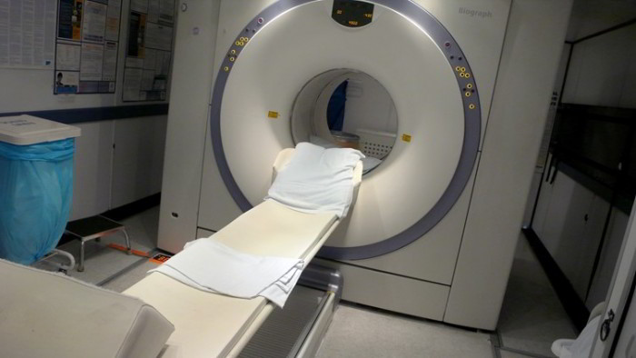
Computerized tomography (CT) scan uses X rays in conjunction with a computer. A string of X rays is passed through the body at slightly different angles to create highly detailed cross sectional images (slices) of the body, named tomograms. CT scan makes it possible to collect comprehensive information about organs painlessly and so that your exposure to radiation is kept to the very least.
A CT scanner includes an X ray source and an X ray sensor, both of which rotate during the process so they stay opposite each other. CT scan uses X rays otherwise from an average X-ray machine to give a higher quality picture.
A CT scan can reveal elements that are impossible to see using normal X rays, including fibrous tissue in solid organs which include the liver.
The advice from the X-ray detector is sent to the computer, which builds up cross- sectional pictures of the body and displays them on a computer screen. These resultant pictures can be saved either as computer files or on traditional X-ray film. More advanced computers can generate three dimensional pictures from conventional CT information. Newer CT scanners use the spiral (or helical) technique, at which at which scanner rotates around you as
A CT scan is a painless process which could take less than one minute to one hour to perform, determined by the state being investigated.
The bed moves forwards slowly so the X ray columns follow a spiral path. This kind of CT scan creates three dimensional pictures and reduces the time taken for the scans to be finished.
During a CT scan you’ll lie on a motorized bed that can transfer you into the scanner. The radiographer will request that you lie quite still and hold your breath while each scan is shot. The bed moves forwards a little more after each scan. If you’re really nervous about the process you might be given a light sedative ahead; you might also be encouraged to bring a relative or friend with you if you believe you’d enjoy moral support. The time it takes to have a CT scan is dependent upon the nature of the scan and may vary from several seconds to an hour.
CT scans are most commonly performed on the head and abdomen. Nevertheless, they are able to additionally be used to direct biopsy processes, in which cells or tissue are taken from internal organs for evaluation. Like traditional X rays, CT scan also creates clear pictures of bone. Blood vessels and regions of high the flow of blood, like the lungs, may also be imaged. These pictures may be improved by using a contrast medium (a material which makes a worthless or fluid-filled construction visible on the picture).


 (65 votes, average: 3.72 out of 5)