It can be helpful to think of the digestive system as a tube running through the body with an opening at each end, the mouth at the top and the anus at the bottom. The tube isn’t a neat, constant shape throughout its course. It swells into a muscular storage organ at the stomach where the food you swallow is thoroughly mixed with digestive enzymes that start breaking it up into a form that the body can use.
At the exit from the stomach more essential digestive juices are added from the gallbladder and pancreas. Food particles get smaller and smaller until, in the exquisitely adapted small intestine (small only in diameter, not length) the tiniest fragments of nutrients can pass easily though the intestine wall straight into the bloodstream.
Why is digestion important?
Consider your body as a machine that runs on a fuel called food. Now, the fuel needs to be compatbile enough for the consumption and that’s why digestion is important. It helps your body to acquire nutrients from food and drink to work in perfect order and stay healthy. Proteins, fats, carbohydrates, vitamins , minerals , and water are what we call the nutrients. Your digestive system breaks nutrients into parts small enough for your body to absorb and use for energy, growth, and cell repair.
– Proteins break into amino acids
– Fats break into fatty acids and glycerol
– Carbohydrates break into simple sugars
The Digestive Process
Human beings take food through mouth and digest it in specific organs for digestion, the undigested food is defecated. The food take passes through a specific canal which begins with buccal cavity and ends at the anus. This canal is called elementary canal or the digestive tract.
The path of the elementary canal are buccal cavity, esophagus, stomach, large intestine, small intestine, rectum and anus. The food gets digested gradually as it passes through the various parts of the track. Digestive juices secreted by the salivary glands liver and pancreas also aid digestion.
Food intake in human beings is through mouth. This process of taking food is called ingestion. Chewing food is very necessary for proper digestion as digestion process starts in the mouth itself.
What happens during chewing?
The large pieces of food we eat are mechanically broken down into small pieces teeth helps us in doing so action of saliva on food during chewing food mixes with saliva. Saliva is secreted from salivary glands located on the lower jaw between the tongue and teeth. Saliva converts starch in the food into sugars and also soft ins the food tongue.
Taste of food can be detected by the taste buds present on the tongue. Tongue is a fleshy muscular organ attached to the floor of the buccal cavity. It is free at the front and moves in all directions. Taste buds appear as small bumps on the tongue and have different regions which help us enjoy different tastes.
The swallowed food from the mouth passes on to the Esophagus. The epiglottis which is a flap like structure behind the tongue directs the food into the Esophagus and prevents entry into the larynx the peristaltic movements of the esophagus helps the food to move down the track till it reaches the stomach.
Stomach is a bag like structure with thick wall it is in the shape of a flattened u and is the widest part in the elementary canal. The food that enters the stomach is churned and mixes with the mucus hydrochloric acid and digestive juices secreted by the inner lining of the stomach. The acid helps to kill the bacteria present in the food. It also makes the medium in the stomach acid and the digestive juice is secreted by the stomach break down proteins into simpler substances then the food is emptied into small intestine.
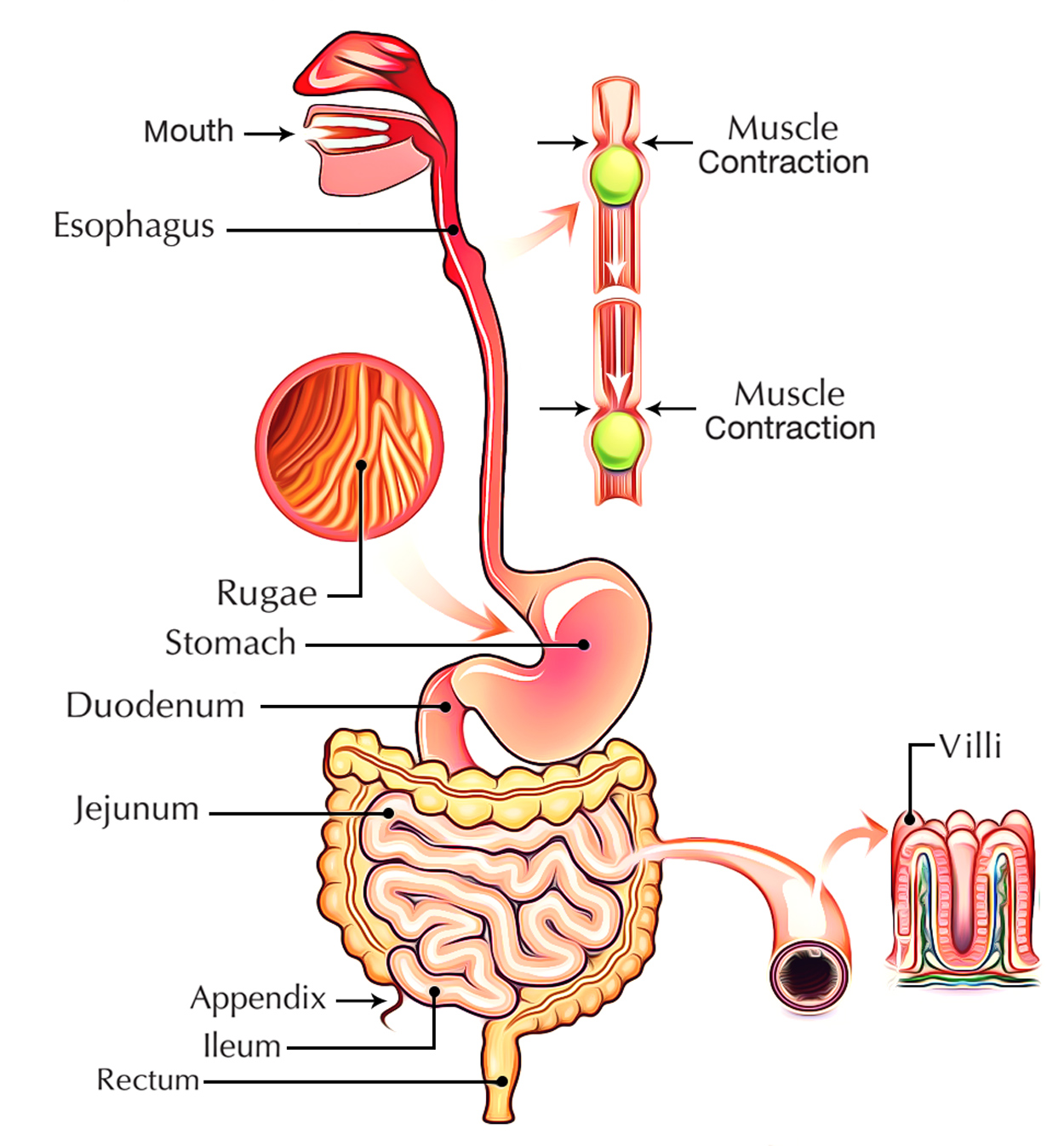
Digestive Process
Small intestine is a highly coiled structure and is about 7.5 meters long as soon as a small intestine receives food from the stomach the digestive juices from liver and pancreas are mixed into it liver.
Liver is the largest gland in the human body and is situated in the upper side of the abdomen to the right it is reddish-brown in color. It secretes bile which breaks down fats bile is stored in gall bladder between meals pancreas pancreas is a leaf shaped and green colored gland located just below the stomach. It secretes pancreatic juice which acts and breaks down carbohydrates proteins and fats after this was completed the partially digested food reaches the lower part of the small intestine where intestinal juices act upon the food and completes digestion of any remaining components in food.
The carbohydrates in the food break down into sugars like glucose fats into fatty acids and glycerol and proteins into amino acids. The food consumed is digested and is now ready to be absorbed into the blood.
Absorption of food in the small intestine the inner wall of the small intestine has finger-like projections called villi which have a network of pin blood vessels close to the surface the villi’s increases the surface area of absorption and absorbs the digested food materials. The substances thus absorbed are transported through the bloodstream to different organs in the body and are used to build complex substances like proteins, this process is called assimilation. In the cells, glucose breaks down into carbon dioxide and water with the help of oxygen and releases energy.
The undigested and unabsorbed food enters the large intestine. Large intestine the large intestine is wider than small intestine and measures 1.5 meters in length once the undigested food enters the large intestine it absorbs water and some salts from it and passes the remaining waste into the rectum here it remains as semi-solid foetus the fecal matter is removed through the anus this is called ejection.
Organs
Pharynx
Stretching from the base of the skull to the esophagus, the pharynx is a funnel-shaped fibromuscular tube. It’s lined throughout with mucous membrane. For both food (deglutition) and air (respiration,) the pharynx functions as a mutual channel. It’s situated behind the cavities of nose, mouth and the larynx with which it interacts.
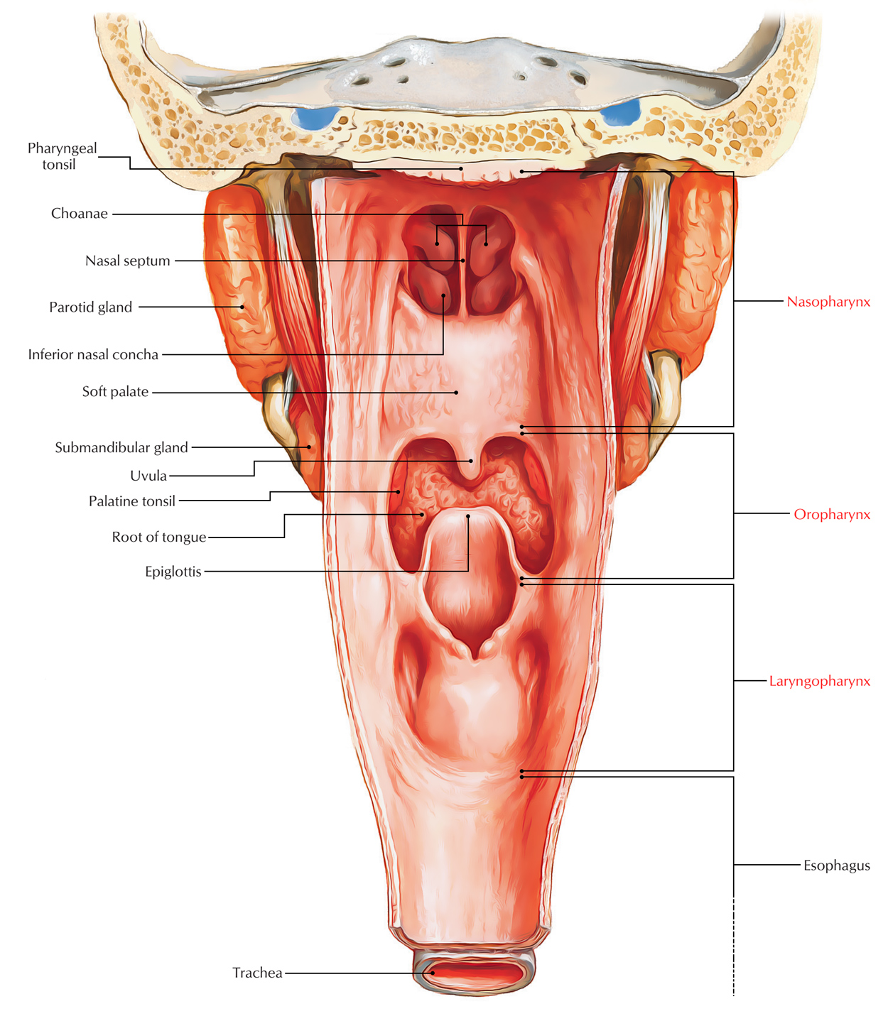
Digestive System: Pharynx
Esophagus
Esophagus is a narrow muscular tube extending from pharynx to the stomach. It gives passage for chewed food (bolus) and liquids during the third stage of deglutition and is about 25 cm long. Its engagement in different diseases like esophagitis, esophageal varices and cancer makes the anatomy of esophagus medically significant. It begins with lower part of the neck and ends in the upper part of the abdomen by joining the upper end of the stomach.
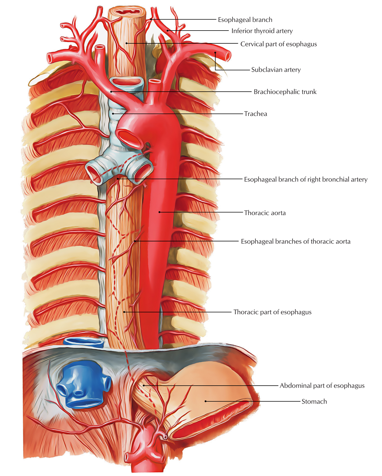
Digestive System: Esophagus
The esophagus begins in the neck at the lower border of the cricoid cartilage (at the lower border of C6 vertebra), descends in front of the vertebral column goes through superior and posterior mediastina, pierces diaphragm in the level of T10 vertebra and ends in the abdomen in the cardiac orifice of the stomach in the level of T11 vertebra.
Stomach
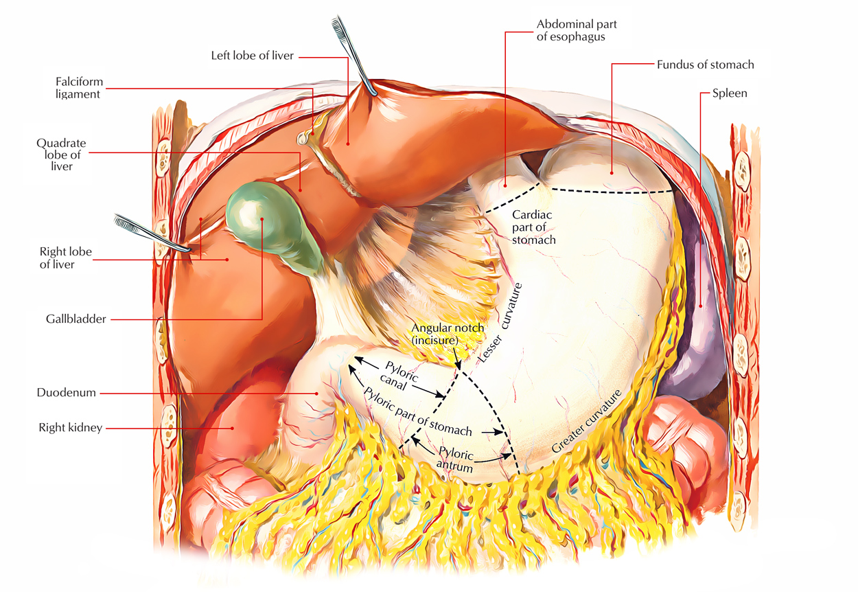
Digestive System: Stomach
The stomach is the part of the alimentary canal between the esophagus and the duodenum. It is the widest and most distensible part. The stomach inhabits the left hypochondriac, umbilical, and epigastric regions. It is located in the upper left part of the abdomen. It flows from the left hypochondriac region into the epigastric region obliquely. The stomach is largely “J” shaped. Its long axis enters downward, forward, and to the right and eventually backward and somewhat upward. It tapers from the fundus on the left of the median plane to the narrow pylorus somewhat to the right of the median plane.
Small Intestine
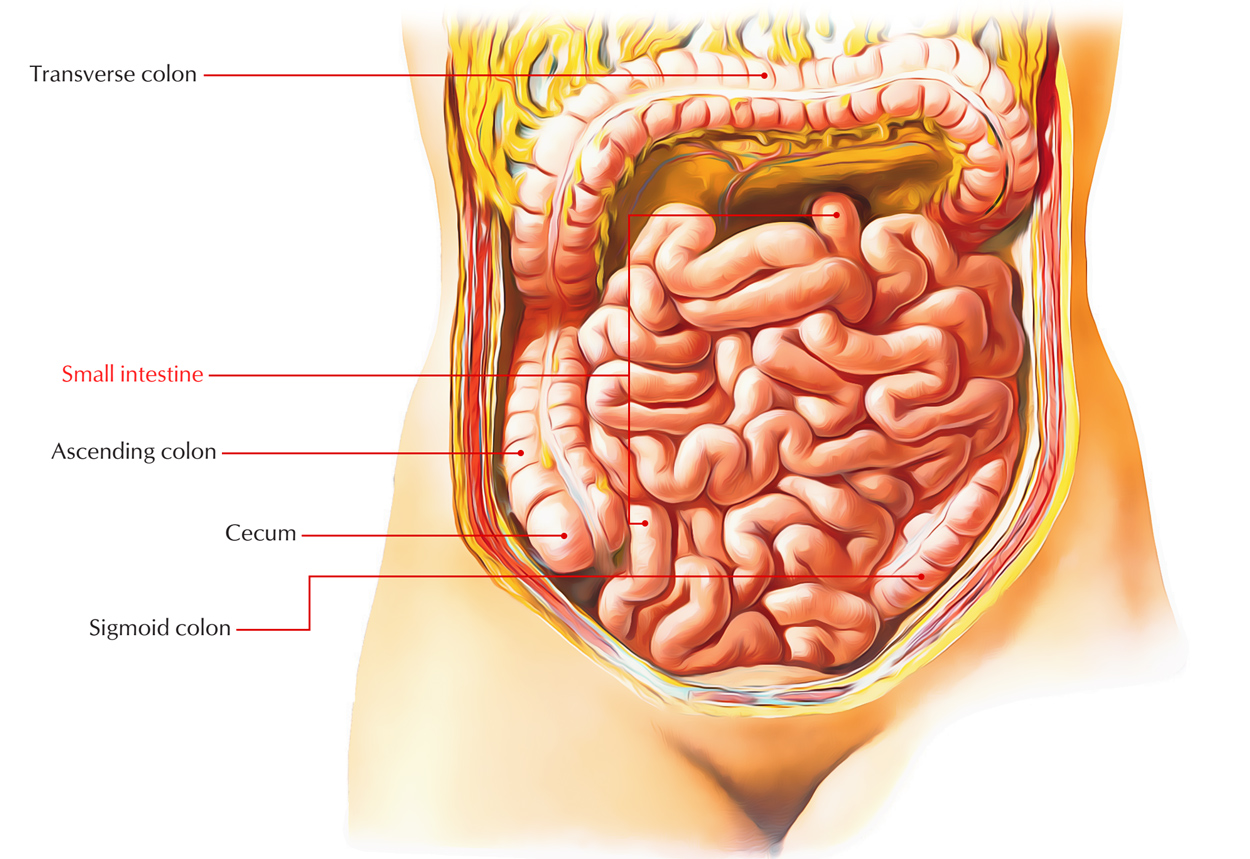
Digestive System: Small Intestine
The small Intestine is a winding, tightly-folded tube in the digestive system that absorbs about 90% of the nutrients from the food we eat. Anatomy says, the small intestine extends from the pylorus to the ileocaecal junction. Its length goes about 6 m and is also divided into 3 parts: duodenum, jejunum, and ileum. The duodenum is the proximal fixed part that is about 10 inches (25 cm) long. The other 2 parts, i.e., jejunum and ileum are freely mobile and stays long.
Spleen
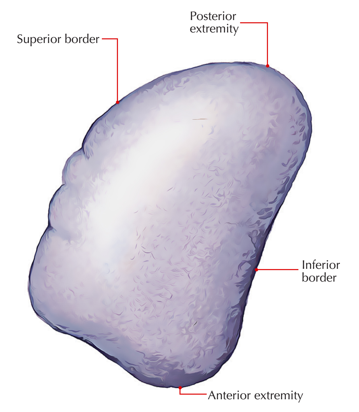
Digestive System: Spleen
Spleen is the largest lymphoid organ in the body (strictly speaking a hemolymphoid organ). The spleen located in the left hypochondrium between the fundus of the stomach and the diaphragm, behind the midaxillary lineopposite the 9th, 10th, and 11th ribs. Its long axis is located parallel to the long axis of the 10th rib. During respiration, it moves a bit from its position.
Liver
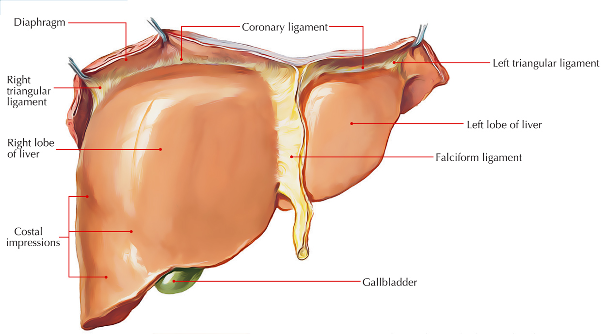
Digestive System: Liver
Liver (Greek hepar. liver) is the largest gland of the body, inhabiting much of the right upper part of the abdominal cavity. Just like in pancreas, the liver is also composed of both exocrine and endocrine parts. The liver performs a broad range of metabolic processes essential for homeostasis, nourishment, and immune response.
Gallbladder
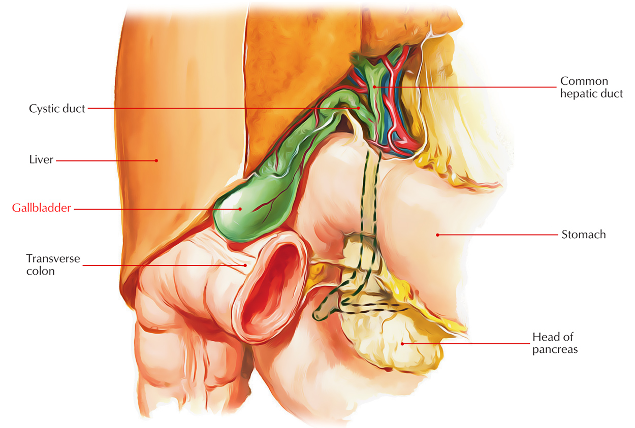
Digestive System: Gallbladder
The Gallbladder is an elongated pear-shaped sac, with about 30-50 ml of capacity. It does the work of storing and concentrating the bile and eliminating it in the duodenum by its muscular contraction. Certain Radiopaque materials that are excreted in the bile are also concentrated in it. To show radiographically (cholecystography) the cavity of gallbladder and its ability to concentrate and contract, they are utilized.
Large Intestine
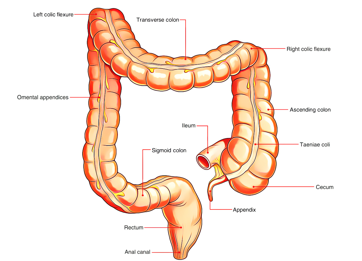
Digestive System: Large Intestine
The Large Intestine is amde up of caecum, colon, rectum and anus. The length of the large intestine goes about 1.5 m. It starts from the caecum to its right, iliac fossa to the anus in the perineum. As compared to the small intestine, it is more fixed in position, aside from the transverse colon and sigmoid colon.
Rectum
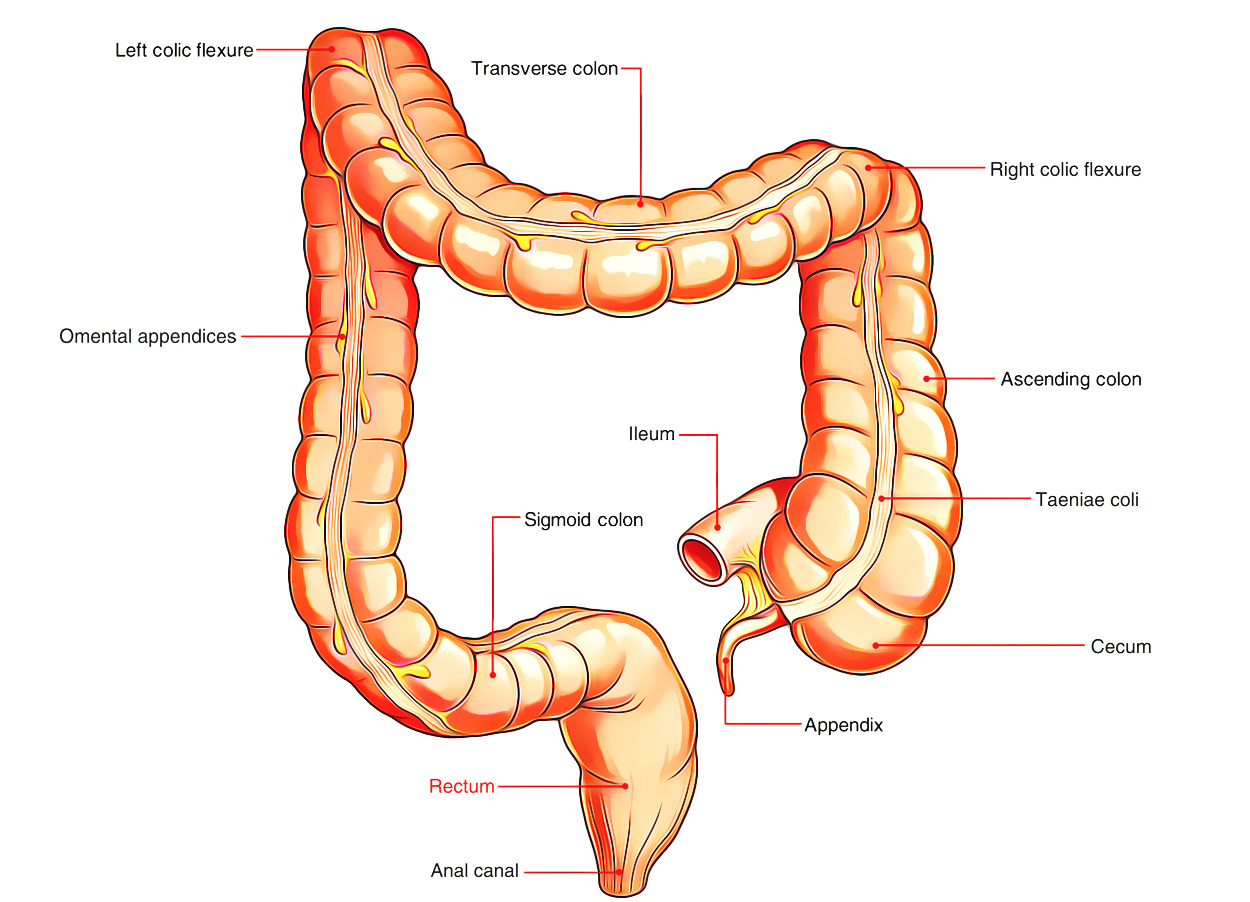
Digestive System: Rectum
The rectum is the distal part of the large intestine between the sigmoid colon and the anal canal. In Latin, the word “rectum” means straight; but the rectum is straight in quadrupeds and not in men. In spite of the fact that the rectum is a part of the large intestine, it’s devoid of taenia coli, sacculations and appendices epiploicae- the cardinal features of the large intestine.
Anal Canal
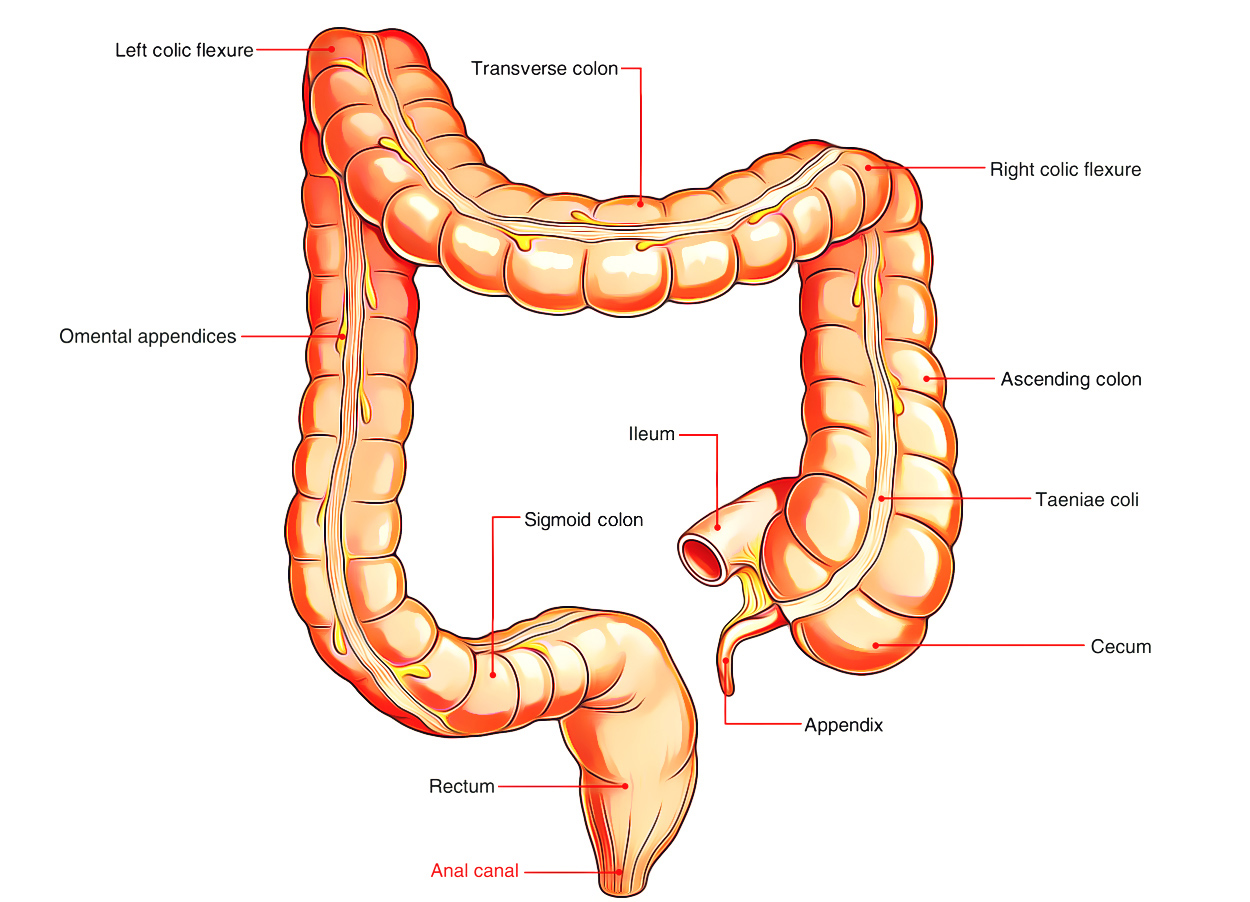
Digestive System: Anal Canal
The Anal Canal is the terminal part (3.8 cm long) of the large intestine, situated in the perineum below the pelvic diaphragm. Like rectum it’s devoid of sacculations, taenia coli, and appendices epiploicae. It’s encompassed by an inner automatic sphincter and an outer voluntary sphincter, whose tone keeps the anal canal closed with the exception of during the defecation.
The anal canal is situated in the anal triangle of the perineum between the left and right fat filled up ischiorectal fossae. These fossae enable the growth of the anal canal during defecation.
Clinical Significance
Many bowel problems revolve around the production of excess acid (ulcers, indigestion and heartburn), the upward escape of acid (hiatus hernia) and disordered movements of the intestine (constipation, diarrhoea and irritable bowel syndrome). The rapid turnover of cells within the digestive tract predisposes to cancer, especially of the stomach and large intestine.
Large Intestine
We hear a lot. about the large intestine (large in diameter) or colon, or more familiar still, the bowel, because it seems to be the focus of many common complaints. Its function in the body is really very simple, it’s there to ensure that precious nutrients and water aren’t lost. Its main job is to absorb water from its fluid contents. When the stream of digested food leaves the small intestine it ’s liquid, very runny. Wren we pass it as a stool it’s solid. Its passage through the large intestine has brought about this change, purely through the absorption of water.
Rectum
The rectum too can absorb water from faeces, in fact, it will wring faeces dry if they stay in the rectum too long. So if the “call to stool” is ignored for any length of time faeces can become stony hard and difficult, even painful, to pass.
This form of constipation is often thought of as a fault of the bowel or diet, but it isn’t. It’s simply due to not emptying the rectum soon enough. And it can be cured by heeding the call to stool as soon as you’re aware of it, even though the pressure to ignore it and do something else is strong. Laxatives aren’t necessary to cure this type of constipation, only retraining yourself to heed what your bowel is telling you to do. If you don’t, your bowel won’t bother to send you the signal and your “constipation” will worsen.
Oesophageal Cancer and Bowel Cancer
If I were to try to pull out the headlines of what we’ve discovered over the last decade two topics would immediately spring to mind, both of them cancer, oesophageal cancer and bowel cancer. The first is referable to heartburn. For decades heartburn has been thought of as nothing more than a mild irritation, a troublesome form of indigestion. In the last live or so years, however, we’ve come to realize that recurrent heartburn, especially if it’s left untreated, can eventually result in malignant change of the lower end of the gullet where it enters the stomach.
It’s caused by the chronic irritation from acid gastric contents, which regurgitate into the lower part of the oesophagus. Here, the lining has no protection from the burning effect of stomach acid. By contrast the stomach lining is covered with a thick protective layer of mucus, so acid never reaches the delicate walls of the stomach lining.
Rigorous treatment of heartburn can prevent cancerous change, so you must seek advice from your doctor if you have a long-term problem.
We know more and more about bowel cancer. In the UK it kills approximately 18,000 people every year and it’s almost certainly due to die fact that our Western diet now contains too little fibre (mainly fruit and vegetables). We can help the next generation by making sure our children eat the magic number of five fruits a day.
Signs of Bowel Cancer
There are two cardinal signs of bowel cancer. The first is seeing blood in your stool, though that’s a rather late sign. Before that you’ll probably have noticed a change in your bowel habits, going more or less often than you used to, a touch of constipation or diarrhoea where previously you had none. Or a sudden change in the shape of the stool narrow where previously it had been round. This change in the bowel habit, in middle age or later, is a sign you must heed and discuss with your doctor.
Peptic ulcer now has a new cause. We used to think it was due to too much stomach acid. We now know it’s due to an infection, a new infection, with Helicobacter pylori. Treatment has been revolutionised in the last 10 years.
Now antibiotics are the Ivnchpin and, as the bacterium is difficult to eradicate, the course lasts a minimum of two weeks, along with other medicines to increase the effectiveness of the antibiotic. One course of treatment and nearly everyone is cured! Now, that’s an advance on the old days.

 (69 votes, average: 4.17 out of 5)