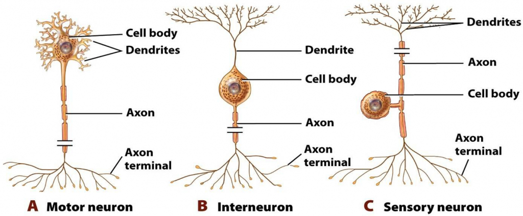
Neurons come in numerous sizes. For instance, a single sensory neuron from your fingertip has an axon that spreads out the length of your arm, while neurons within the brain may extend just a couple of millimeters. They also have different shapes depending upon their functions. Motor neurons that regulate muscle contractions have a cell body on one point, a long axon in the middle and dendrites on the other point. Sensory neurons have dendrites on both ends, connected with a long axon with a cell body in the middle. Interneurons, or associative neurons, bring information between motor and sensory neurons.

Motor, Sensory and Interneuron
Sensory System
Receptors
The initialization of sensation stems from the action of a specific receptor to a physical stimulus. The receptors which react to the stimulus and start the procedure of sensation are commonly defined in four distinct categories: chemoreceptors, photoreceptors, mechanoreceptors, and thermoreceptors. All receptors receive unique physical stimuli and transduce the signal into an electrical action capacity. This action potential then travels along afferent neurons to specific brain areas where it is processed and translated.
Chemoreceptors
- Chemoreceptors, or chemosensors, detect specific chemical stimuli and transduce that signal into an electrical action capacity. The two primary types of chemoreceptors are:
- Distance chemoreceptors are essential to receiving stimuli in the olfactory system through both olfactory receptor neurons and neurons in the vomeronasal organ.
- Direct chemoreceptors consist of the taste buds in the gustatory system as well as receptors in the aortic bodies which identify alterations in oxygen concentration.
Photoreceptors
- Photoreceptors are capable of phototransduction, a method which converts light (electromagnetic radiation) into, other kinds of energy, a membrane capacity.
- The three primary kinds of photoreceptors are: Cones are photoreceptors which respond substantially to color. In human beings the three different kinds of cones refer a primary reaction to short wavelength (blue), medium wavelength (green), and long wavelength (yellow/red).
- Rods are photoreceptors which are really sensitive to the intensity of light, enabling vision in dim lighting. The concentrations and ratio of rods to cones is highly associated with whether an animal is diurnal or nocturnal.
- In human beings rods outnumber cones by around 20:1, while in nighttime animals, such as the tawny owl, the ratio is more detailed to 1000:1. Ganglion Cells live in the adrenal medulla and retina where they are involved in the sympathetic response. Of the ~ 1.3 million ganglion cells present in the retina, 1-2% are said to be photosensitive ganglia. These photosensitive ganglia contribute in vision for some animals, and are believed to do the same in human beings.
Mechanoreceptors
Mechanoreceptors are sensory receptors which react to mechanical forces, such as pressure or distortion. While mechanoreceptors exist in hair cells and play an essential function in the vestibular and auditory systems, most of mechanoreceptors are cutaneous and are grouped into four categories:
- Slowly adapting type 1 receptors have small responsive fields and react to static stimulation. These receptors are mostly used in the sensations of form and roughness.
- Slowly adapting type 2 receptors have large receptive fields and react to extend. Similarly to type 1, they produce sustained reactions to an ongoing stimulus.
- Rapidly adjusting receptors have small receptive fields and underlie the understanding of slip.
- Pacinian receptors have large receptive fields and are the predominant receptors for high-frequency vibration.
Thermoreceptors
- Thermoreceptors are sensory receptors which react to varying temperature levels. While at the same time the systems through which these receptors run is uncertain, recent discoveries have shown that mammals have at least two distinct kinds of thermoreceptors:
- The end-bulb of Krause, or bulboid corpuscle, detects temperatures above body temperature.
- Ruffini’s end organ identifies temperatures below body temperature level.
Nociceptors
Nociceptors respond to possibly destructive stimuli by sending signals to the spinal cord and brain. This procedure, called nociception, generally causes the understanding of pain. They are found in internal organs, and also on the surface of the body. Nociceptors detect various type of harmful stimuli or actual damage. Those that only respond when tissues are damaged are called “sleeping” or “quiet” nociceptors.
- Thermal nociceptors are activated by poisonous heat or cold at numerous temperature levels.
- Mechanical nociceptors respond to excess pressure or mechanical contortion.
- Chemical nociceptors respond to a wide array of chemicals, some of which are indications of tissue damage. They are associated with the detection of some spices in food.
Motor-System
- The sensory systems create our mental images of the external world. These representations offer us with details and cues that guide the motor systems to create movements produced by the collaborated contractions and relaxations.
- The motor systems are hierarchically organized in the central nervous system (CNS) as the spinal neuronal circuits that control the automatic stereotypic reflexes.
- Higher centers in the brainstem moderate postural regulated and balanced locomotor movements. The highest centers, consisting of the motor areas of the cerebral cortex, initiate and regulate intricate skilled voluntary movements.
The major components of the somatic motor system are arranged and longitudinally oriented along the neuraxis as two path systems:
(1) The phylogenetically new direct pathways that fine-tune and control voluntary movements namely the corticospinal tract and the corticobulbar tract coming from the cerebral cortex and project to end in the anterior horn of the spinal cord and nuclei of the brainstem.
(2) The phylogenetically old more scattered and indirect pathways that primarily mediate reflex and postural regulation of the musculature, namely coming down indirect pathways from the cortex to brainstem nuclei that protrude to the anterior horn of the spinal cord (e.g. corticoreticular and cortico preticospinal tracts).

 (51 votes, average: 4.57 out of 5)