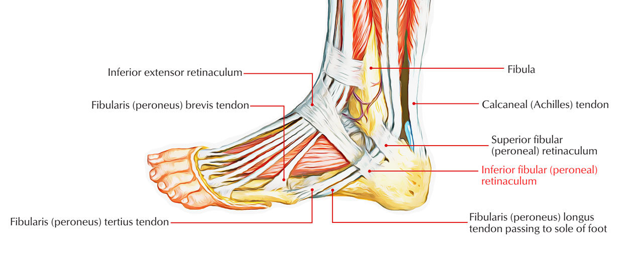
The inferior peroneal retinaculum is an extension of the lateral inception of the inferior extensor retinaculum. It has origin via the posterolateral edge of the sinus tarsi along with each of the superficial as well as deep fibers. Superficial fibers route plan- tarly as well as posteriorly, go across the peroneal trochlea, as well as enter superior to the calcaneal tubercle, while deep fibers enter over the trochlear procedure. This structure creates two fibrous tunnels (inferior peroneal tunnel) above the trochlear procedure, a superior tunnel for the peroneus brevis along with an inferior tunnel for the peroneus longus.

Inferior Peroneal Retinaculum
Attachments
- Superiorly: It is connected to the anterior part of superior side of calcancum, near to the branch of inferior extensor retinaculum.
- Inferiorly: It is connected to the lateral side of calcancum.
- Between: It is connected to the peroneal trochlea, hence creating two loops, one for the tendon of peroncus brevis along with another for the tendon of peroncus longus.
Relations
The tendon of peroneus brevis goes through the superior loop which of peroneus longus via the inferior loop of the inferior retinaculum. Every single tendon is confined in a different synovial sheath that is the extension of the common synovial sheath above, below the superior peroneal retinaculum.
Clinical Significance
The synovial sheaths confining the tendons of peroneus longus along with peroneus brevis undergo abrasion as well as swelling in professional athletes who use tight shoes.

 (51 votes, average: 4.60 out of 5)